Sunday, September 12, 2010

Tissues!

Here are pictures of the slides that the tutor Randy set up in open lab Sunday.
I have labeled them the same as the blue paper stated.

This is Adipose Connective Tissue with the pointer at an adipocyte


Areoler connective tissue

Dense Irregular tissue (not from our slides)


Dense Regular Tissue with pointer at fibroblast

Same as above, but closer    
Elastic Cartilage
Fibrocartilage with pointer at chondrocyte
Hyaline Cartilage
Pointer at Cilia
Psuedo-stratified columnar ciliated
Reticular with pointer at a reticular protein fiber (small thing black strand)
Simple Columnar
Simple columnar (up close)
Simple Cuboidal (up close)
Simple Cuboidal
Simple Squamous Cells
Stratified Squamous Keratinized
Transitional 
That is all I have, please feel free to post your own pictures in comments, labeled. I will let Mrs. Jaurez know so she's able to check and make sure all labels are correct! Goodluck this week!

Tuesday, August 31, 2010

Lab one: Torso Models, Animal/Human Cell Models, Skin Model, Brain Model, Ear Model, Muscle Leg Model, Kidney Model

My torso model pictures are on my phone which is currently dead, so here are the other models. Please look for an update tomorrow with the torso model, thanks :)

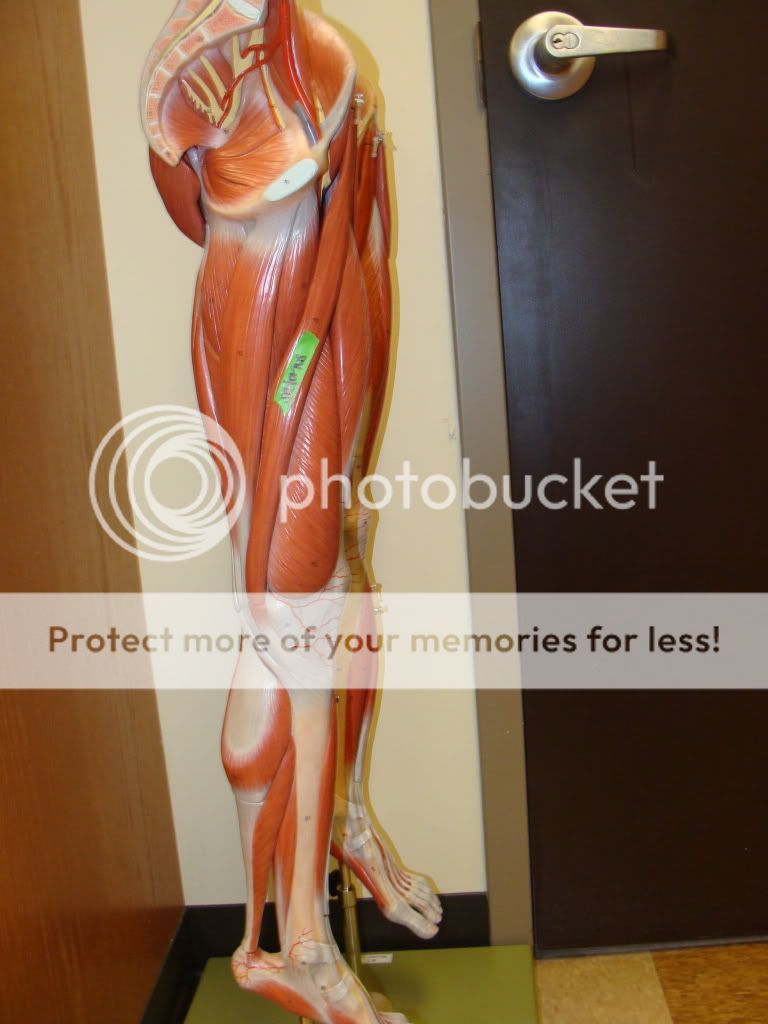
These pictures show different angels of the Muscle leg model #M-2
Labeled is the:
Sartorius Muscle


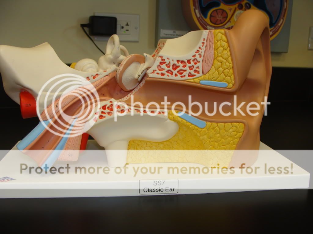
Here is the ear model #SS-7
with the external acoustic meatus labeled.


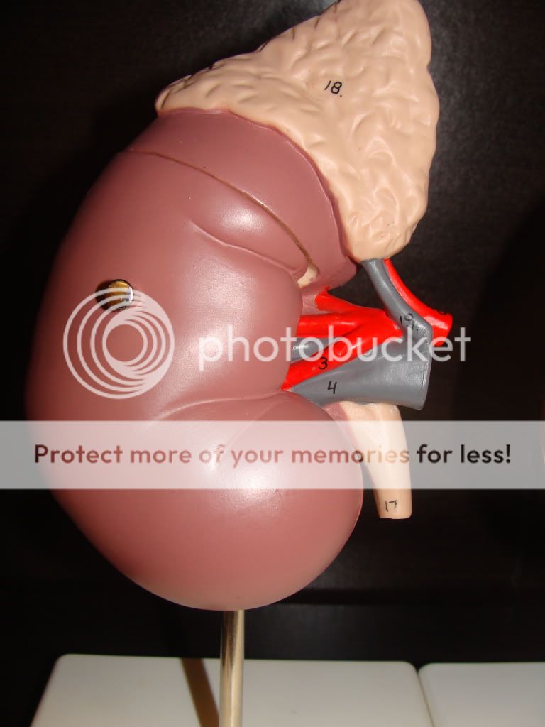
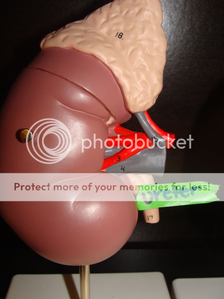
Kidney model # U-1 with the ureter labeled


Cervical vertebrae model #N-4 with the subarachnoid cavity labeled

Sorry I didn't get a great picture but if you look at the model it's on the right side. :)

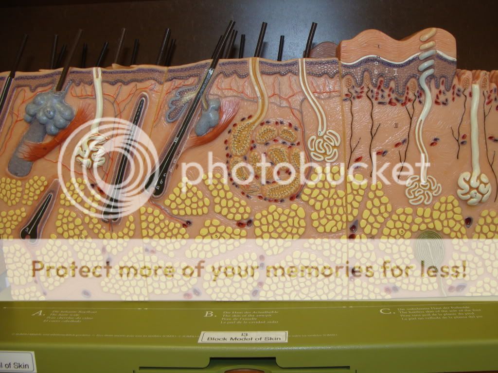
This is skin model #1-3 and the Meissner's Corpuscle is labeled


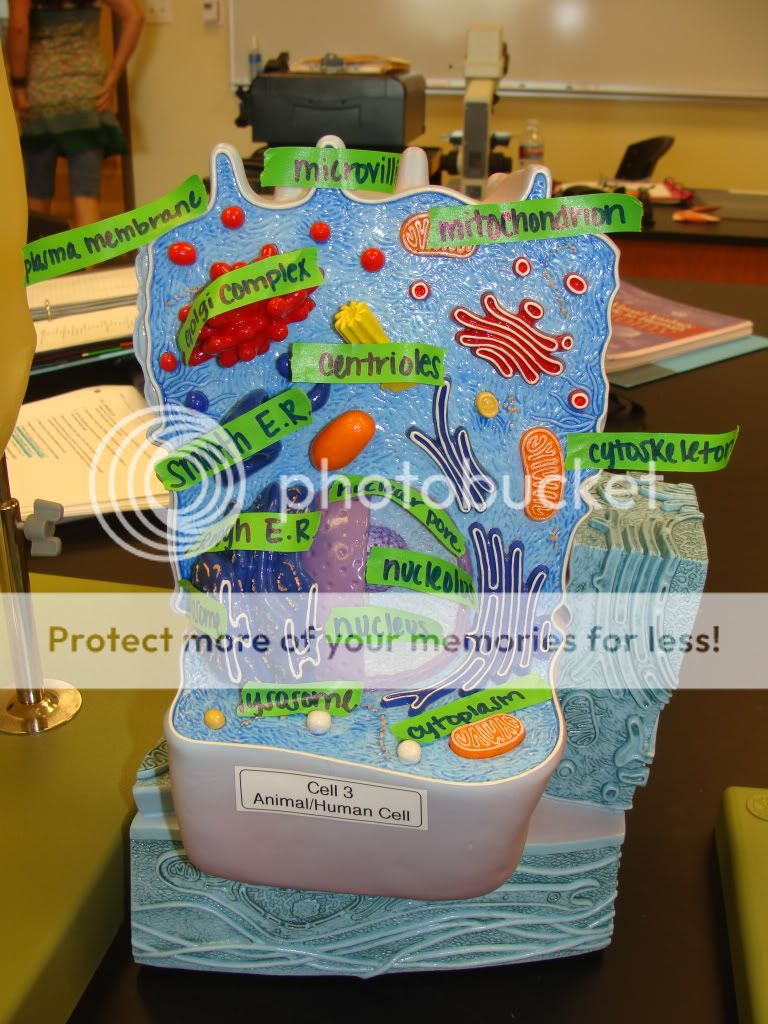
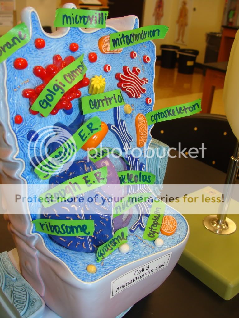
Here are different human/animal cells
the following labels are:
Smooth Endoplasmic Reticulum
Rough Endoplasmic Reticulum
lysosome
golgi complex
centrioles
microvilli
plasma membrane
nuclear pores
ribosomes
cytoskeleton
cytoplasm
nucleus
nucleolus
mitochondrion
cilia

Please note that the above model didn't have visable cytoskeleton, cilia, or microvilli.


Again, If you need torso model pictures check back tomorrow and they will be on here.
Hope this helps anyone who couldn't go to open lab

Saturday, August 28, 2010

Hello There

Hey Anatomy Friends:

 I'm Nataly,  and I designed this blog to help people study for their practical exams and labs. As you know we will be taking lots of pictures to help us learn parts of the body... So that's what I'm doing... taking my pictures and posting them on here after class so that all of you are able to come to the site and use my pictures to assist you into getting that "A" that we all want :).

I will be taking these with a professional camera so that our pictures can be clear as day. If you'd like to send me a picture that you'd like me to add, send it to nataly.metcalfe@austincc.edu.

As you noticed I do not have pictures from the first lab, so if anyone would like to send those to me I'll get started with putting a detailed description of everything we need to know. I will also use this blog for open questions through comments.. so feel free to ask anything.. we can learn so much from working together.

Lastly, I will put study material that I have found useful and we can also set up study groups/dates on here.

I hope this is useful to everyone in our class

-Nataly